
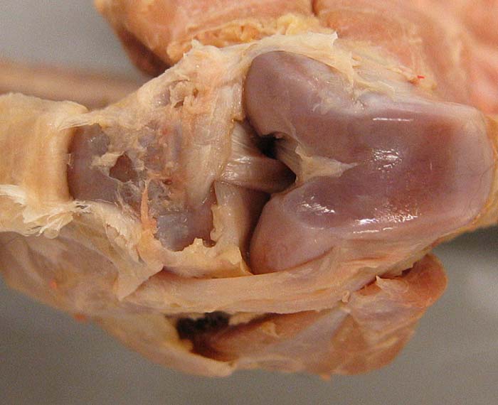
Lab 7 - Image 18
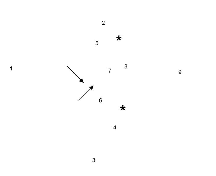
Feline stifle joint dissection, cranial view: The joint capsule has been removed and the patellar ligament (1) is reflected distally. The joint is extremely flexed. Medial (2) and lateral (3) collateral ligaments are evident. The tendon of origin (4) of the long digital extensor m. passes through the joint to reach the lateral femoral condyle. Medial (5) and lateral (6) menisci are bound to the tibia by ligaments (arrows). A cranial cruciate ligament (7) and a caudal cruciate ligament (8) can be seen between femoral condyles (asterisks). Notice the trochlea (9) of the femur.