
Lab 7 - Image 11
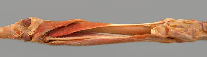

This deep caudal dissection in the cat reveals that two deep muscles remain following removal of the gastrocnemius m., soleus m., superficial digital flexor m., and lateral & medial heads of the deep digital flexor m. The two deep muscles of the caudal crus are the popliteus m. (1) and the caudal tibial m. (2). The latter is a tarsal extensor muscle that is relatively better developed in the cat than in the dog. The tendon of origin of the popliteus m. can be seen passing deep to the lateral collateral ligament of the stifle joint. Notice the tibia (asterisk) and fibula (arrow). (The fibularis brevis m. (3) of the craniolateral muscle group is evident.)