
Lab 6 - Image 5
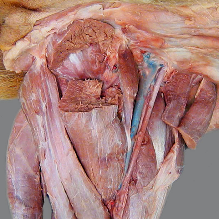
Ventral view of the pelvis and medial view of the thigh: The external obturator m. (1) is exposed by transecting the adductor m. (2), which originates from the symphysial tendon (3).
Caudally, notice the two bellies of the semimembranosus m. (4) and the displaced semitendinosus m. (5). Cranially, femoral triangle (6) is bounded by the pectineus m (7) and the two parts of the sartorius m. (8), which has been transected. A portion of tensor fasciae latae m. (9) is evident cranial to the quadriceps femoris m. (which is not labeled).