
Lab 6 - Image 18
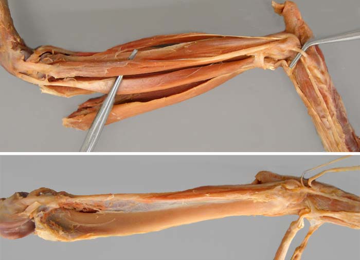
Cat dissection: Top: Lateral view of the crus and pes (caudal muscles removed). The fibularis longus m. (1) and its tendon are isolated by probes. The fibularis brevis m. (2) is exposed by displacement of the slender lateral digital extensor m. (3). The long digital extensor m. (4) and the detached cranial tibial m. (5) are bound distally by crural extensor retinaculum (6). Also, tarsal extensor retinaculum (7) is evident.
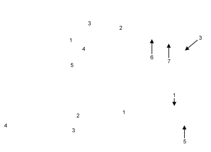
Bottom: Lateral view of the crus and tarsus with most of the muscles removed. The fibularis brevis m. (1) originates from the fibula (2). The tibia (3) and femur (4) are exposed. Notice that tarsal extensor retinaculum (5) is preserved.