
Lab 6 - Image 11
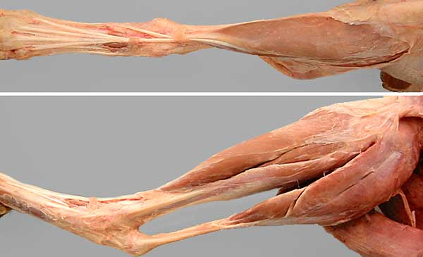
Top: Cranial view of the crus and dorsal view of the pes. The tendon of the cranial tibial m. (1) curves medially at the tarsus to insert on the plantar surface of the metatarsus. The tendon of the long digital extensor m. (2) branches in the metatarsus. Extensor retinaculum consists of two components: the crural extensor retinaculum (3) and, more distally, the tarsal extensor retinaculum (4)
Bottom: Lateral view of the crus and pes. Identify these craniolateral-group muscles: cranial tibial m. (5), long digital extensor m. (6), and fibularis longus m. (7). (The caudal group of crus muscles includes: deep digital flexor m. (8), superficial digital flexor m. (9), and gastrocnemius m. (10).) Notice the semitendinosus m. (asterisk).