
Lab 5 - Image 4
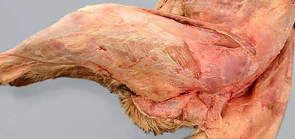
Removal of the subcutaneous, loose areolar, superficial fascia, uncovers a membranous deep fascia along the back, rump, tail, thigh and crus. The deep fascia sheets are named according to their regional location: thoracolumbar deep fascia (1); gluteal deep fascia (2); fascia lata (3); and crural fascia (4). (Caudal fascia of the tail is obscured by reflected skin in this specimen.)
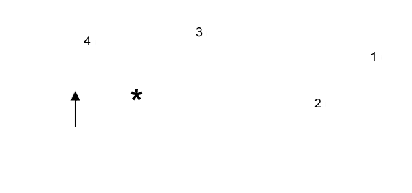
The politeal lymph node (asterisk) is embedded in loose areolar deep fascia caudal to the stifle joint. (The lateral saphenous vein (arrow) emerges distal to the lymph node.)