
Lab 4 - Image 15
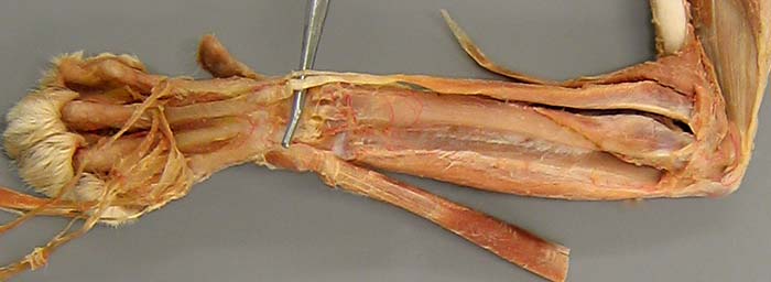
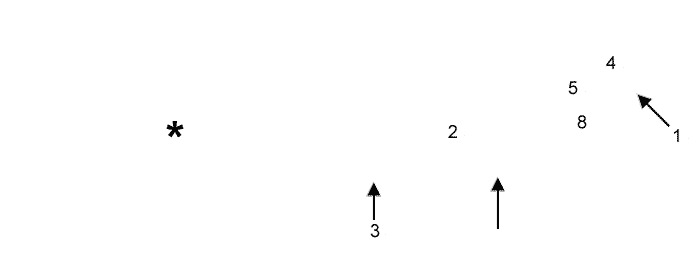
Cat, left antebrachium, lateral view. Most of the musculature has been transected and reflected away. The cut edge of the common digital extensor m. (1) is evident. The pronator quadratus m. (2) is exposed, between the radius and ulna. The reflected extensor carpi ulnaris (ulnaris lateralis) m. (3) is labeled. A probe has elevated tendons of the extensor carpi radialis longus (4) and extensor carpi radialis brevis (5) mm. (the longus tendon is artifactually detached). The supinator m. (8) is labeled. An arrow points to the flexor carpi ulnaris m., a caudal muscle exposed by the dissection. The asterisk indicates a metacarpal bone.