
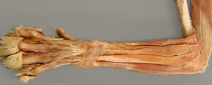
Cat, left antebrachium, lateral (cranio-lateral) view. Deep fascia has been removed from the antebrachium and metacarpus, leaving extensor retinaculum (asterisk) binding tendons at the carpus. Distal to the retinaculum, notice the branched tendon of the common digital extensor m. (1). The tendon of the lateral digital extensor m. (2) also passes deep to the retinaculum. The most lateral muscle is the extensor carpi ulnaris m., also named ulnaris lateralis m. (3). In the cat, the extensor carpi radialis m. has two distinct parts, named: extensor carpi radialis longus (4) and extensor carpi radialis brevis (5). The anconeus m. (6) is visible proximally.
Typically the cat has a well developed brachioradialis m., but that muscle is missing (arrow) in this specimen. In the brachium, the long head of the triceps brachii m. is preserved.