
Lab 3 - Image 9
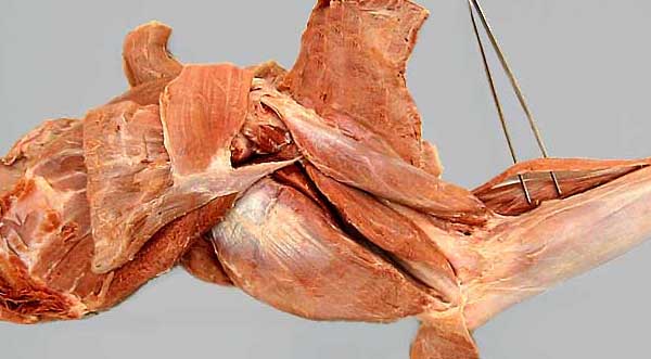
Medial view, with the tensor fasciae antebrachii m. (1) transected and reflected: Identify the long (2), accessory (3), and medial (4) heads of the triceps brachii m., the biceps brachii m. (5), and coracobrachialis m. (6). Also evident are: teres major m. (7), subscapularis m. (8), latissimus dorsi m. (9), omotransversarius m. (10), and pectoral mm. (11).
In the antebrachium, forceps elevate the brachioradialis m. (which is better developed in the cat than the dog).