
Lab 3 - Image 7
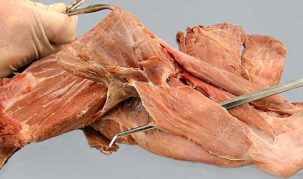
Medial surface of the brachium, caudal view: A probe has been placed deep to the thin tensor faciae antebrachii m. (1). Its tendon attaches to the latissimus dorsi m. (2) which is being pulled cranially (an attachment of cutaneus trunci m. (3) is evident). Identify: long (4) and medial (5) heads of the triceps brachii m., the biceps brachii m.(6), and the attachments of superficial pectoral and cleidobrachialis mm. (asterisks). On the scapula, the teres major m. (7) and the subscapularis m. (8) can be seen.