
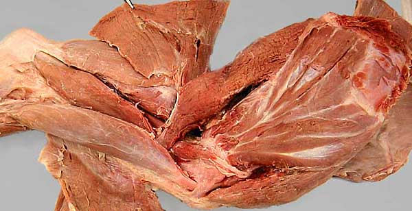
On the medial surface of the scapular, notice the: teres major m. (1), the subscapularis m. (2), and a portion of the supraspinatus m. (3). The small coracobrachialis m. (4), with its long tendon of origin, can be seen. An asterisk marks the area (serrated face) where the serratus ventralis m. had attached.
On the medial surface of the brachium, notice the: biceps brachii m. (5). Its tendon of origin passes deep to the transverse humeral retinaculum (arrow). Medial (6), accessory (7), and long (8) heads of the triceps brachii m. can be seen. The tendon of origin of the tensor faciae antebrachii m.(9) joins the tendon of the latissimus dorsi m. (10), which in turn joins the tendon of the teres major m. Pectoral muscles (11) attach to the cranial surface of the humerus (12).