
Lab 2 - Image 4
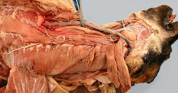
Lateral view of the ventral neck (dog). The external jugular vein (1) is reflected medially to expose the esophagus (2) on the left side. Contents of the carotid sheath [common carotid a., etc.] (3) are barely evident dorsal to (below) the esophagus.
Muscles visible in this advanced dissection include: serratus ventralis m. (4); omotransversarius m. (5); cleidomastoid part of the brachiocephalicus m.(6); and the sternocephalicus m. (7).
[Also evident are the three parts of the scalenus m. (asterisks); rectus throacis m.(8) and fascicles of cervical intertransversarius mm. (9)]