
Lab 2 - Image 11
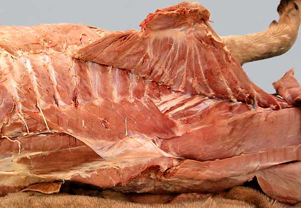
The top of the scapula is pulled laterally to reveal the serratus ventralis m., which has a limb attachment medially on the scapula and a broad trunk attachment on ribs and cervical vertebrae. Thus the serratus ventralis m. has cervical (1) and thoracic (2) divisions. The serratus dorsalis m. (3), which is not a limb muscle, is also exposed. Attachments of both serratus muscles have a serrated appearance. Notice the rhomboideus m. (4). The latissimus dorsi m. has been removed. Epaxial (back) muscles are marked with asterisks.