Hind Limb: Femoral Fracture
RETURN
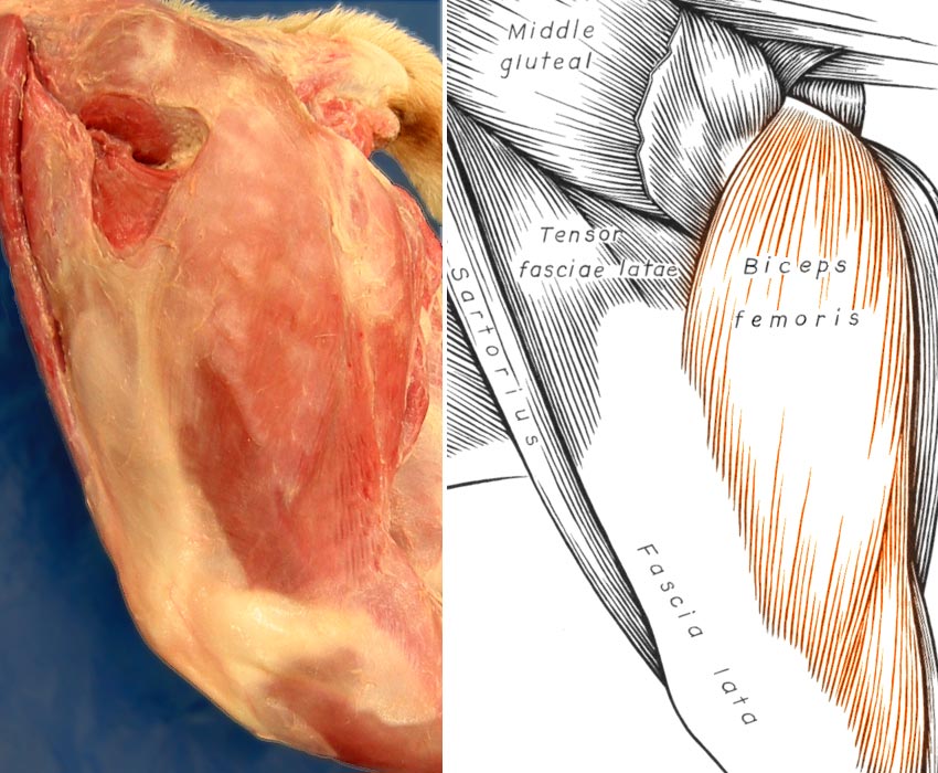
In this composite image, a superficial dissetion of the canine thigh is shown on the left and a schematic drawing of muscles is shown on the right. The biceps femoris muscle is colored. The positions of the biceps femoris muscle and fascia lata should be noted. In the craniolateral surgical approach to the femur, an incision is made just cranial to the biceps femoris muscle through the relatively avascular fascia lata.
Go Top