
Close
Caudal Mesenteric Ganglion & Hypogastric Nerves
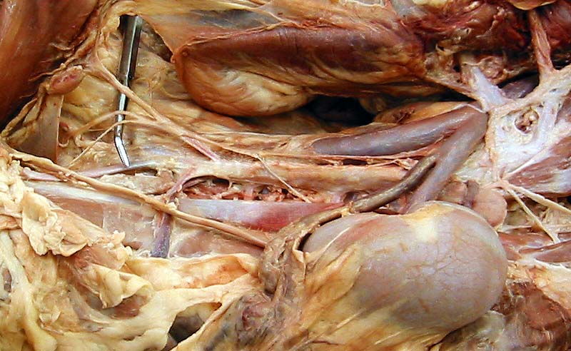
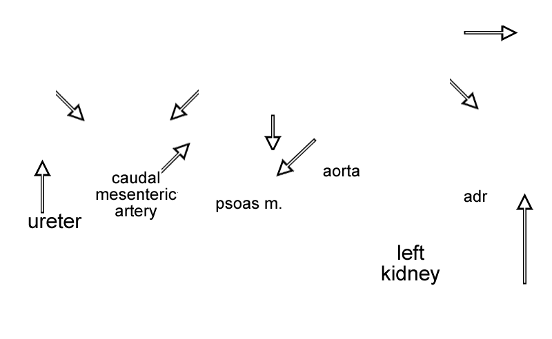
Left ventral view of the dorsal wall of a canine abdomen, showing bilateral hypogastric nerves entering the pelvic canal. The nerves arise from the caudal mesenteric ganglion located within the caudal mesenteric nerve plexus which runs on the caudal mesenteric artery. Plexuses on the caudal mesenteric artery and on the aorta receive preganglionic sympathetic axons from typically four lumbar splanchnic nerves (one is preserved in the illustrated dissection).
Cranially, major (maj splanch) and minor splanchnic nerves convey preganglionic axons to the adrenal gland (adr) and to the celiac ganglion and the cranial mesenteric ganglion (cran mesenteric gang). Also labeled are psoas minor muscle (psoas m.), aorta, celiac artery, kidney, ureter, and descending colon.
Cranially, major (maj splanch) and minor splanchnic nerves convey preganglionic axons to the adrenal gland (adr) and to the celiac ganglion and the cranial mesenteric ganglion (cran mesenteric gang). Also labeled are psoas minor muscle (psoas m.), aorta, celiac artery, kidney, ureter, and descending colon.
[spacebar toggles labels — esc closes window]