| Home Page |
General ANS Features |
Sympathetic Division |
Parasympathetic Division |
Trace Pathways |
Physiology & Pharmacology |
The term Enteric Nervous System refers to autonomic neurons that collectively innervate the alimentary tract within which they reside. The expansive volume of the alimentary tract, the great number of neurons located within the gut wall, and the relative independence of gut reflex activity from the CNS control all make autonomic innervation of the alimentary tract a special case, compared to innervation of other viscera.
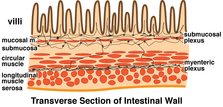
Within the gut wall, autonomic nerves are found in two locations: The submucosal (Meisner) plexus is located within the submucosa. The myenteric (Auerbach) plexus is found between circular & longitudinal muscle coats:

Each plexuses contains:
• preganglionic axons from the vagus nerve;
• postganglionic neurons of parasympathetic terminal ganglia;
• axons from postganglionic neurons located in sympathetic prevertebral ganglia; and
• innervation from the pelvic plexus (for descending colon, rectum, & anal canal)
• axons of visceral afferent neurons (conveying mechanoreceptor & chemoreceptor activity)
Sympathetic and parasympathetic innervation originating from the central nevous system (CNS) plays an important role in controlling alimentary tract function. However, a remarkable amount of gut reflex activity occurs independently of the CNS. Visceral afferent nerves can trigger: local reflexes by synapsing on postganglionic neurons within submucosal or myenteric plexuses or regional reflexes by synapsing on postganglionic neurons in prevertebral autonomic ganglia.
