

Quick Links
Web-Based Instruction
Veterinary Planar Anatomy
(Arranged Categorically)
To view a web site click its title.
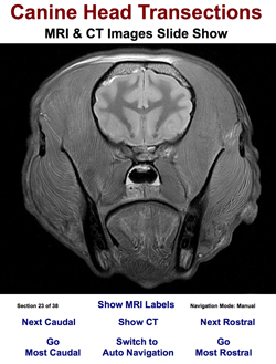
Canine Head Transections (MRI & CT Slide Show)
This web app presents a slide show of MRI and CT axial (transverse plane) images through the head of an embalmed, latex-injected, brachycephalic, mongrel canine cadaver. The web app enables students to explore planar head anatomy thoroughly and to compare MRI vs CT views at each transverse level. MRI and CT images are synced during rostral-caudal navigation, which can be manual or automatic (at an adjustable rate). During manual navigation, MRI labels may be displayed (using a button or the spacebar). The keyboard can be used for navigation (arrow keys) and to toggle MRI/CT display (shift key). The auto navigation mode gives students an overview of head planar anatomy. The MRI labels that become available during manual navigation provide detailed identification.

Canine MRI Atlas
This web site presents MRI images of the canine head, neck, thorax, abdomen (male & female), and pelvis (male & female). Each region can be viewed in transverse, sagittal & dorsal planes; each image has accompanying labels that can be toggled on/off. The mouse or keyboard may be used to navigate through the images and toggle the appearance of labels. At all times, single-key navigation executed by Return/Enter is available: regions [h / n / t / a / p], planes [x / s / d], gender [m / f], and location within an image set [1...9 (10...90%)]. The MRI images are proton-density-weighted from embalmed, latex injected cadavers.
Canine Head MRI Atlases
This web site presents images of a cadaver Beagle Head (Cranium) obtained by Magnetic Resonance Imaging (T2-weighted images from a 3 Tesla MR system). Three Atlases show MRI images in transverse, sagittal, and dorsal planes of view. Buttons toggle labels and switch between bright and dark versions of the images, a slider generates gradations between bright and dark image versions.

Canine Regional Planar Anatomy Quizzes
This web site enables students to explore canine planar anatomy. MRI, CT and Cadaver-Slab images, displayed in Transverse Plane, Dorsal Plane and Sagittal Plane views, are presented in random order per body region. Two modes of presentation are available: In Quiz Mode, three terms and five target frames are shown per image. Students match terms to frames by dragging or sequentially clicking/tapping. In Atlas Mode, numerous labels per image may be toggled on/off.

MRI Regional Quiz Puzzles
NEW. This web app invites students to regard MRI quizzes as puzzles to be solved: using anatomical knowledge obtained by cadaver dissection to interpret structural relationships evident in planar MRIs. Students are challenged to maximize percent correct scores while completing 12 answers per MRI. As shown to the right, users click/tap a circle beside an anatomical term and then a target circle on the image. When correct, a red line will connect the two red circles. When wrong, the percent correct score decreases and one must choose again. (Previous successful choices are indicated by gray and black colors.)
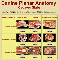
Canine Cadaver Planar Anatomy (cadaver slabs)
Canine planar anatomy is presented as 900 x 600 pixel images of cadaver slabs produced by bandsawing frozen cadavers. The head/neck, thorax, and abdomen/pelvis regions are each shown in three planes: sagittal (left to right lateral views), transverse (cranial/rostral to caudal, caudal views), and dorsal (dorsal to ventral, dorsal views).
Keyboard arrow keys may be used to navigate among images per plane per region. Use the spacebar or click/tap an image to toggle labels.

Canine Brain MRI Atlas
This web site presents labeled transverse views of a Beagle Brain obtained by Magnetic Resonance Imaging: T2-weighted, T1-weighted, and Proton-Density-weighted MRIs are included. Three viewing options are presented: 1] MRIs are paired with stained tissue sections to facilitate neuroanatomy identification; 2] brain MRIs are shown in situ within transverse views of the Beagle head; and 3] MRIs are featured in animated interactive quizzes intended to offer knowledge self-assessment of MRI brain anatomy (click/tap a term and then its corresponding target; if correct, a connecting line will appear).

mobile MRI Brain Atlas
This web app presents proton-density-weighted MRI images paired with brain-transection images of a dog brain. The app is intended to provide quick access to canine brain MRI anatomy through a smart phone. Particular brain levels are accessible via a Gallery Menu (tap a small image) or a List Menu (tap a text button). At each level, a button toggles between MRI and brain section images. Labels per image can be switched on/off by tapping the image (or a Show/Hide button). Rostral/caudal buttons are available for navigation to adjacent levels. This web app can be viewed on a smart phone, a tablet, or a computer screen.

Canine Brain Atlas
This Brain Atlas web site consists of twelve transverse levels through a dog brain. The Atlas is accompanied by a Glossary-Index of brain anatomy terms. Labels may be toggled on/off and levels may be navigated with a mouse or key strokes. (Page width is 1200 pixels; a print version of this Atlas is available in A Practical Guide to Canine & Feline Neurology, 2nd ed. by Curtis W. Dewey, Wiley-Blackwell 2008.)
Additional Instructional Material & Links
Link: University of Illinois Radiographic Anatomy Imaging
The University of Illinois has produced an outstanding web site for viewing normal radiographic anatomy images. The canine is most thoroughly presented, followed by feline, equine and bovine images. The site is still undergoing development. Your browser must activate a Flash Player to view this web site.